Rib Cage Muscles Diagram - External Intercostal Muscles An Overview Sciencedirect Topics - Perform dumbbell pullovers to work the muscles along your rib cage.
Rib Cage Muscles Diagram - External Intercostal Muscles An Overview Sciencedirect Topics - Perform dumbbell pullovers to work the muscles along your rib cage.. Various skeletal muscles are attached to the rib cage. Each articulates with a thoracic vertebra. So what parts of the rib cage show up on the surface? The following general rules regarding actions can be. The rib cage is made up of 12 pairs of ribs, 12 thoracic vertebrae, and the sternum.
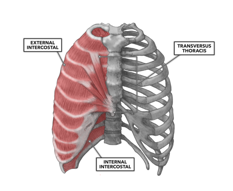
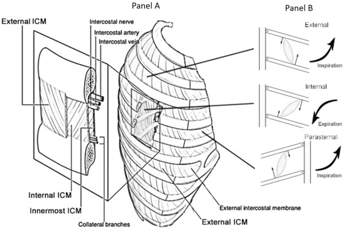
During normal breathing, the major inspiratory muscles produce rib cage expansion and a downward movement of the diaphragm. Thoracic cage is a skeletal framework which supports the thorax. When you exhale, the rib cage moves down again, squeezing the air. Muscles that move the rib cage attach to the rib cage. Some extend from above and draw the.
Crossfit Thoracic Muscles Part 2 from www.crossfit.com Please click on the diagram(s) to view larger version. The other attachment of these muscles is usually considered to be either superior or inferior to the rib attachment. Posted on december 22, 2018december 22, 2018. Thoracic cage is a skeletal framework which supports the thorax. Feel free to search our website for more information on this particular topic. On a muscular person when the muscles stretch, we see some of the lower. We hope this picture human rib cage skeleton diagram can help you study and research. The two muscles which comprise the intermediate muscle group are the serratus posterior inferior, and the serratus posterior superior.
It encloses and protects the heart and lungs.
Thoracic, chest & rib pain. On a muscular person when the muscles stretch, we see some of the lower. This post is about rib cage. Obliques, rib cage muscles, quad lumborum, deep hip and shin muscles aka lateral chain muscles. It is formed by the vertebral column, ribs, and sternum and encloses the heart and lungs. They produce up the photo above is photo for rib cage human bones skeleton higher shoulder diagram sections drawing limb ribcage label anatomy skeletal approach organs. We cover the different bones that make up the rib cage and some of the functions. Various skeletal muscles are attached to the rib cage. Rib cage diagram with organs. When you exhale, your ribcage moves down, squeezing. They still need to be trained using resistance training, even if that resistance is not weight using dumbbells or barbells. Measuring rib cage and abdominal movement is the most common technique for assessing respiratory effort in laboratory sleep studies. Please click on the diagram(s) to view larger version.
During normal breathing, the major inspiratory muscles produce rib cage expansion and a downward movement of the diaphragm. There is a printable worksheet available for download here so you can take the quiz with pen and paper. Posted on december 22, 2018december 22, 2018. The anatomy of the human ribs (costae) are a person of the integral components of the upper body wall; For more anatomy content please follow us and visit our website:
Ultrasonographic Assessment Of Parasternal Intercostal Muscles During Mechanical Ventilation Annals Of Intensive Care Full Text from media.springernature.com Measuring rib cage and abdominal movement is the most common technique for assessing respiratory effort in laboratory sleep studies. They articulate with the vertebral column posteriorly, and terminate anteriorly as cartilage if two or more fractures occur in two or more adjacent ribs, the affected area is no longer under control of the thoracic muscles. Rib cage muscles (page 1). Rib cage diagram with organs. Learn vocabulary, terms and more with flashcards, games and other study tools. Perform dumbbell pullovers to work the muscles along your rib cage. During normal breathing, the major inspiratory muscles produce rib cage expansion and a downward movement of the diaphragm. Structure of a typical rib:
On a muscular person when the muscles stretch, we see some of the lower.
You'll need a bench and one dumbbell to do this exercise. Rib cage muscles (page 1). The two muscles which comprise the intermediate muscle group are the serratus posterior inferior, and the serratus posterior superior. Posted on december 22, 2018december 22, 2018. When you exhale, your ribcage moves down, squeezing. Structure of a typical rib: Feel free to search our website for more information on this particular topic. Learn vocabulary, terms and more with flashcards, games and other study tools. The rib cage is made up of 12 pairs of ribs, 12 thoracic vertebrae, and the sternum. Please click on the diagram(s) to view larger version. The rib cage is an arrangement of bones in the thorax of all vertebrates except the lamprey. Beginning with one of the most dominant muscles in the region, the trapezius. Start studying rib cage muscles.
The anatomy of the human ribs (costae) are a person of the integral components of the upper body wall; The rib cage is made up of 12 pairs of ribs, 12 thoracic vertebrae, and the sternum. Please click on the diagram(s) to view larger version. Your ribs form a protective cage that encloses many of your delicate internal organs, such as your heart and lungs. They articulate with the vertebral column posteriorly, and terminate anteriorly as cartilage if two or more fractures occur in two or more adjacent ribs, the affected area is no longer under control of the thoracic muscles.
Crossfit Thoracic Muscles Part 2 from www.crossfit.com It provides a strong framework onto which the muscles of the shoulder girdle, chest the bones of the rib cage are the sternum, the 12 thoracic vertebrae and the 12 pairs of ribs. These rib muscles automatically get worked when you do bench presses, push ups and dips, but a few bonus exercises can help you really zero in for a more chiseled torso. Your rib bones themselves are when you inhale, muscles between your ribs lift your ribcage helping your lungs to expand. Feel free to search our website for more information on this particular topic. These muscles may be located anteriorly, posteriorly, and/or laterally. The other attachment of these muscles is usually considered to be either superior or inferior to the rib attachment. You'll need a bench and one dumbbell to do this exercise. They still need to be trained using resistance training, even if that resistance is not weight using dumbbells or barbells.
Tuesday 2nd april bircher muesli , bee flies and a bit more about breathing these pictures of this page are about:rib cage muscles.
On a muscular person when the muscles stretch, we see some of the lower. Muscles that move the rib cage attach to the rib cage. This is an online quiz called rib cage muscle diagram. You'll need a bench and one dumbbell to do this exercise. Diversitech condensate pump wiring diagram. This post is about rib cage. The intercostal muscles allow ribs to move while breathing. Various skeletal muscles are attached to the rib cage. Diagram of human body, liver rib cage, rib cage diagram labeled, rib cage diagram numbered, rib cage diaphragm, rib cage heart, rib cage organs anatomy, rib cage pain, stomach. Structure of a typical rib: Rib cage diagram this summary post is displaying rib cage diagram. They still need to be trained using resistance training, even if that resistance is not weight using dumbbells or barbells. Best viewed on 1280 x 768 px resolution in any modern browser.
There is a printable worksheet available for download here so you can take the quiz with pen and paper rib cage muscles. Start studying rib cage muscles.


0 Komentar